


NANO编辑优选:MiRNA-520a-3p结合叶酸共轭修饰Fe2O3@PDA 多功能纳米试剂用于核磁成像及基因-光热抗肿瘤治疗

文章介绍
Xue Li(历雪), Shuang Wang(王爽), Qingzhe Gao(高庆哲), Na Li(李娜), Shanshan Dong(董珊珊), Yuwei Gao(高玉伟), Zuobin Wang(王作斌), Butian Zhang(张卜天) and Xiuxia He(何秀霞)
通讯作者:
- 何秀霞,长春理工大学
研究背景:
Chemotherapy and radiotherapy combined with surgery are the traditional treatments for many osteosarcoma patients, but the fifive years survival is still less than 60% . Immunotherapy, gene therapy (GT) and targeted therapy have become the preferred choices for adjuvant therapy. Osteosarcoma (OS) is an invasive malignant tumor that originates from the bone, and the majority of osteosarcomas occur in the metaphysis of the distal femur, proximal tibia, and proximal humerus. They are mainly on the surface of the body and visible to the eyes. Malignant proliferating sarcoma cells directly produce tumorous osteoid tissue or immature bone, which are highly gene heterogeneous. Thus, combination therapy strategies are being considered for the treatment of osteosarcoma.
研究内容:
Experimental :
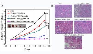
First, γ-Fe2O3 was coated with PDA polymer through a polymerization reaction to obtain Fe2O3@PDA core–shell nanocomposites with high MR imaging capability. Fe2O3@PDA was further modifified with aminoterminated polyethylene glycol (NH2–PEG–NH2) to improve the stability of Fe2O3@PDA in physiological buffer. The PDA shell of Fe2O3@PDA-PEG not only showed a strong photothermal effect, but also offered active sites for loading the therapeutic drugs (miRNA520a-3p) and FA through π–π stacking and hydrogen bond interactions. We prepared FA surface modified miRNA-Fe2O3@PDA-FA nanoparticles that could interact with the corresponding receptors to treat osteosarcoma. The miRNA-Fe2O3@PDA-FA nanodrug can be employed as an effificient nanoplatform for biomedical applications, including MR imaging, highly targeted PTT, and GT against tumors.
Theoretical approach:
(1)Preparation and characterization of the miRNA-Fe2O3@PDA-FA drug delivery system
(2)In vitro biological activity and toxicology investigation of miR-Fe2O3@ PDA-FA
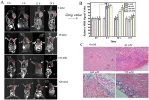
(3) In vivo imaging and toxicology investigation
(4) In vivo MRI imaging
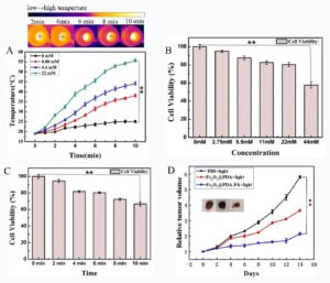
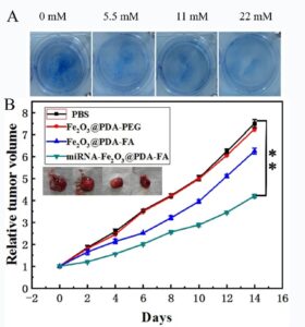
(5)In vivo phtothermal therapy
(6)In vivo GT
(7)In vivo phtothermal/GT
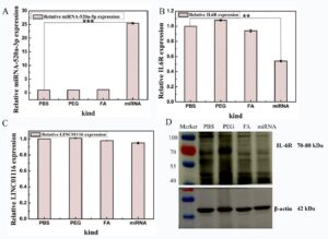
(8)Quantitative real-time polymerase chain reaction (qRT-PCR) and Western blot assay
Results and their significance:
In this study, we evaluated the potential of the multifunctional nanoagent miRNA-Fe2O3@ PDA-FA to deliver the tumor-suppressor miRNA520a-3p for GT for the fifirst time. The nanoagent miRNA-Fe2O3@PDA-FA can make full use of the magnetic properties of Fe2O3 to monitor and diagnose tumor tissues by MRI. The nanoagent miRNA-Fe2O3@PDA-FA can take full advantage of the photothermal properties of PDA. When tumors are exposed to NIR laser, the high temperature produced by miRNA-Fe2O3@PDA-FA can contribute to kill cancer cells. The nanoagent miRNA-Fe2O3@PDA-FA can make the best of the targeted molecular properties of FA, which can effificiently help to deliver miRNA520a-3p to tumor tissues, overexpressed miRNA520a-3p to inhibit tumors, and the FA of miRNA-Fe2O3@PDA-FA can enhance the PTT and GT ofosteosarcoma. Taken together, the fabricated miRNA Fe2O3@PDA-FA acts as an excellent strategy for combination therapy using a ‘one stone that could kill two birds,’and the GT/ PTT/MRI platform has great potential in the future clinical treatment of tumors.
作者介绍

何秀霞 教授
长春理工大学
- He Xiuxia, PhD candidate, Professor, School of Life Science and Technology, Changchun University of Science and Technology. She received a bachelor’s degree in 1998; a master’s degree in Genetics and breeding from PLA Military Supply University in 2003, and a Doctor’s degree in Biochemistry and Molecular biology from Jilin University in 2006. In 2013, she worked at the Analytical Chemistry Postdoctoral Mobile Station of Changchun Institute of Applied Chemistry, Chinese Academy of Sciences. At present, she is an expert in dissertation review at the Academic Degree Center of the Ministry of Education.Her research interests include anti-tumor, anti-bacterial and anti-viral nanodrugs.
期刊介绍
- 2022年影响因子:3.5 Citescore:6.7
- Nanotechnology(NANO)创刊于1990年,是第一本纳米科研和技术领域的专业期刊。NANO发表纳米技术研究发展前沿的高水平研究论文及纳米研究进展的综述,主要集中在纳米能源、生物和医学、电子和光子、图案和纳米加工、传感和驱动、材料合成和材料性能等领域。